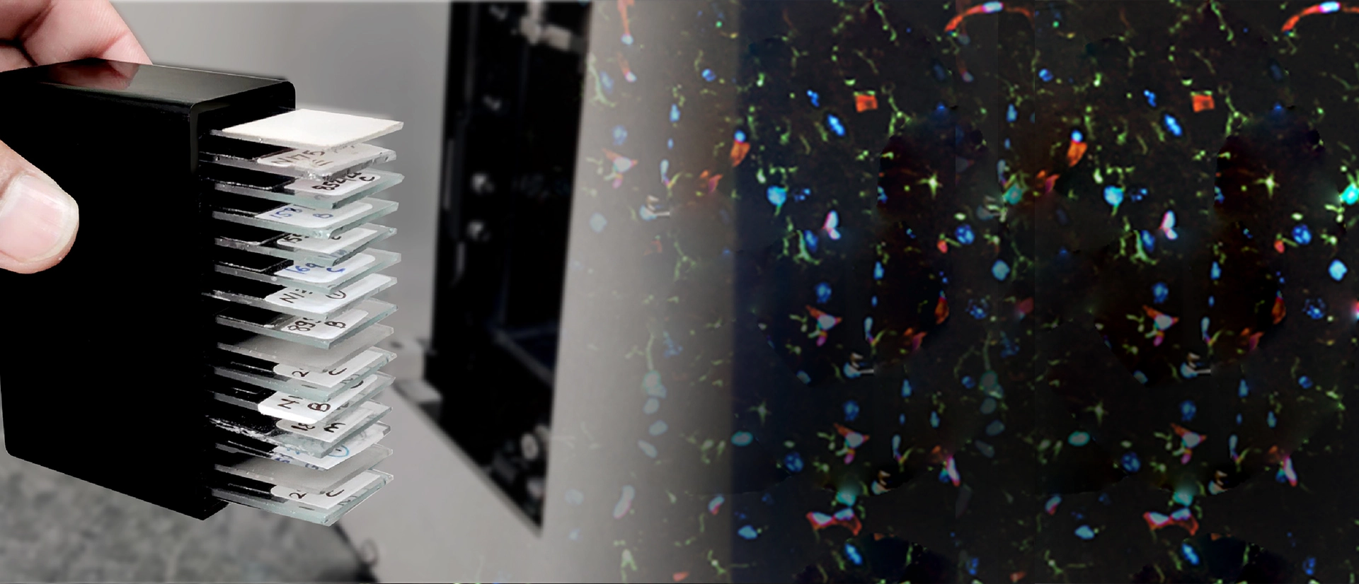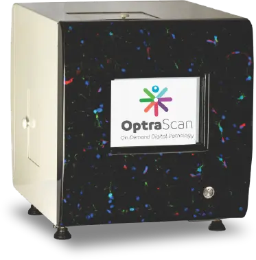

Our OS-FL fluorescence imaging scanner is designed to empower researchers, clinicians, and laboratories with cutting-edge capabilities for a wide range of applications. With unparalleled performance and versatility, OS-FL redefines the standards for fluorescence imaging, delivering exceptional results with every scan.
OptraScan's OS-FL fluorescence imaging scanner is engineered to provide high-resolution imaging across a spectrum of fluorescent dyes and labels. Built with advanced optics and intelligent software algorithms, OS-FL offers precise detection and quantification of fluorescence signals, enabling detailed analysis of cellular structures, biomarkers, and molecular interactions. Whether you're conducting basic research, diagnostic pathology, or drug discovery, OS-FL delivers unmatched imaging quality and reliability.
These scanners come bundled with OptraSCAN's proprietary software solutions such as IMAGEPath® an Image Management System tailored for viewing, storing, and archiving as well as TELEPath®, Telepathology software used for real-time, remote consultations.

A fluorescence slide scanner illuminates tissue samples with specific wavelengths of light, causing fluorophores within the sample to emit light at longer wavelengths, which is then captured by a camera to generate high-resolution images for analysis.
Fluorescence scanning offers superior contrast, enabling visualization of specific cellular structures or molecular markers within samples that may be otherwise difficult to discern with traditional brightfield imaging. Additionally, fluorescence scanning allows for multiplexing, enabling simultaneous detection of multiple targets within the same sample.
Fluorescence slide scanners can image various biological samples including tissue sections, cell cultures, microarrays, and protein arrays, allowing for detailed analysis of cellular structures, protein expression patterns, and molecular interactions. These scanners are particularly useful in biomedical research, pathology, drug discovery, and diagnostics.
Fluorescence scanning enables researchers to study cellular processes, biomarker expression, and disease mechanisms with high sensitivity and specificity, advancing understanding of biological systems and facilitating drug discovery. In diagnostics, it aids in identifying disease markers, characterizing tissues, and guiding treatment decisions with enhanced precision and efficiency.
When selecting a fluorescence slide scanner, factors such as resolution, sensitivity, multiplexing capabilities, compatibility with fluorophores, and software features for image analysis and data management should be evaluated to ensure optimal performance and suitability for specific research or diagnostic needs. Additionally, considerations regarding throughput, automation, cost, and support services may influence the decision-making process.
Yes, fluorescence slide scanners can be integrated with other laboratory equipment such as automated Lab management systems and image analysis software, enabling streamlined workflows and enhanced data analysis capabilities.