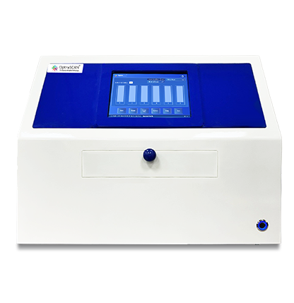

Introducing OS-FS™, OptraSCAN’s Frozen Section Imaging Modality, designed to support rapid intraoperative consultation through real-time digital imaging and remote collaboration. Featuring a Live View Mode, OS-FS™ enables pathologists and hospital networks to review frozen sections as they are prepared, helping deliver timely insights when surgical decisions matter most. By enabling live digital microscopy and seamless connectivity, OS-FS™ supports efficient frozen section workflows without the need for traditional whole slide scanning, reinforcing OptraSCAN’s leadership in real-time digital pathology solutions.
OS-FS™ is a dual-mode frozen section imaging solution that supports both FFPE and frozen section workflows using interchangeable cartridges. Designed for speed-critical environments such as intraoperative pathology, OS-FS™ allows pathologists to instantly review frozen sections or fine needle aspiration (FNA) samples locally or remotely using Live View Mode, reducing waiting times for surgeons and patients. Rather than creating archived whole slide images, OS-FS™ functions as a real-time digital microscope, enabling pathologist-driven review and consultation without workflow disruption. This approach aligns with the unique clinical requirements of frozen section pathology, where immediacy and expert interpretation are essential.
OS-FS™ integrates seamlessly with OptraSCAN’s digital pathology ecosystem, including IMAGEPath and TELEPath.
• IMAGEPath supports image viewing, case organization, and workflow coordination
• TELEPath enables real-time remote consultation and collaboration between pathologists across locations
Together, these tools create a connected frozen section workflow that enhances speed, access, and collaboration, making OptraSCAN a trusted partner for modern pathology departments and hospital networks.
Take a closer look at the future of digital slide imaging and explore the Frozen Section Pathology in our blog.

Intraoperative Diagnostics*: Enable pathologists to rapidly examine tissue samples during surgery, providing real-time diagnostic* information to surgeons for immediate decision-making.
Tumor Identification: Quickly evaluate suspicious lesions or tumors for malignancy, facilitating immediate diagnosis* and treatment planning.
Lymph Node Evaluation: Assist in the intraoperative assessment of lymph nodes for metastatic spread, aiding in accurate cancer staging and informing decisions on lymph node dissection and adjuvant therapy.
Training and Education: Serve as valuable tools for training pathology residents and medical students in interpreting histological specimens and making intraoperative diagnoses*.
Pediatric Surgery: Evaluate tissue samples from pediatric patients during surgery to diagnose congenital abnormalities, tumors, and other conditions requiring prompt intervention, ensuring timely and accurate treatment.
Frozen section pathology involves rapid freezing and sectioning of tissue samples for immediate microscopic examination during surgery, providing quick diagnostic* information, whereas standard histopathology involves formalin fixation, tissue processing, embedding, sectioning, staining, and microscopic examination in a laboratory setting, taking longer for results.
Frozen section pathology is utilized to guide surgical decisions by providing real-time diagnostic* information, especially in scenarios requiring immediate decisions regarding tumor margins, tissue adequacy, or presence of infection.
Biopsy specimens, surgical margins, and lymph nodes are suitable for frozen section analysis.
Frozen section pathology offers rapid diagnosis*, aiding surgeons in making immediate decisions during procedures, while also facilitating timely adjustments to surgical plans based on real-time pathological findings.
Frozen section pathology is typically comparable in accuracy to standard histopathology, with reported sensitivities and specificities ranging from 90-100%, although it may have slightly higher rates of false negatives or false positives due to rapid processing and limited sampling. This is also highly dependent on the quality of the product offered by pathology scanner providers.
Yes, frozen section pathology enables real-time intraoperative decision-making by providing rapid diagnostic* information during surgery, assisting surgeons in determining appropriate next steps regarding tumor margins, tissue adequacy, or presence of infection.