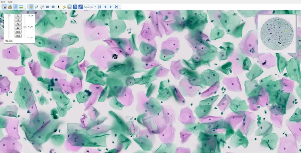
And Extended Depth Of Field Volume Scan Solution For Cytology
Conventional scan routines within a narrow focal range have limitations for Cytology smear slides, as cytology slides have three-dimensional cell groups. This complex multilayer distribution of cells requires multi-layer/multiplanar scanning – a critical component for morphologic analysis for cytopathologists. OptraSCAN’s volume scan routine provides a dual mode for scanning and viewing cytology smears; Z – stacking and extended depth focusing algorithm.
A stack of images is generated at different focal planes along the z- axis. The viewing software enables the user to navigate, zoom up and down the different planes to detect the three-dimensional regions in focus.
Volume scan using z-stacks is expensive in terms of scan time and size due to its multilayer nature. OptraSCAN’s extended depth of field algorithm generates a single, entirely focused composite image to screen an entire image effectively, easily, and systematically.

