A complete digital cytology solution combining brightfield scanning, AI-powered triage, and automated reporting — making cervical cancer screening faster, more consistent, and accessible at any scale.
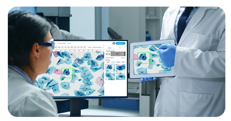
CytoSiA combines brightfield slide scanning, AI-powered cell classification, and automated Bethesda-standardized reporting into a single workflow — helping labs deliver faster, more consistent cervical cancer screening without adding staff or infrastructure.

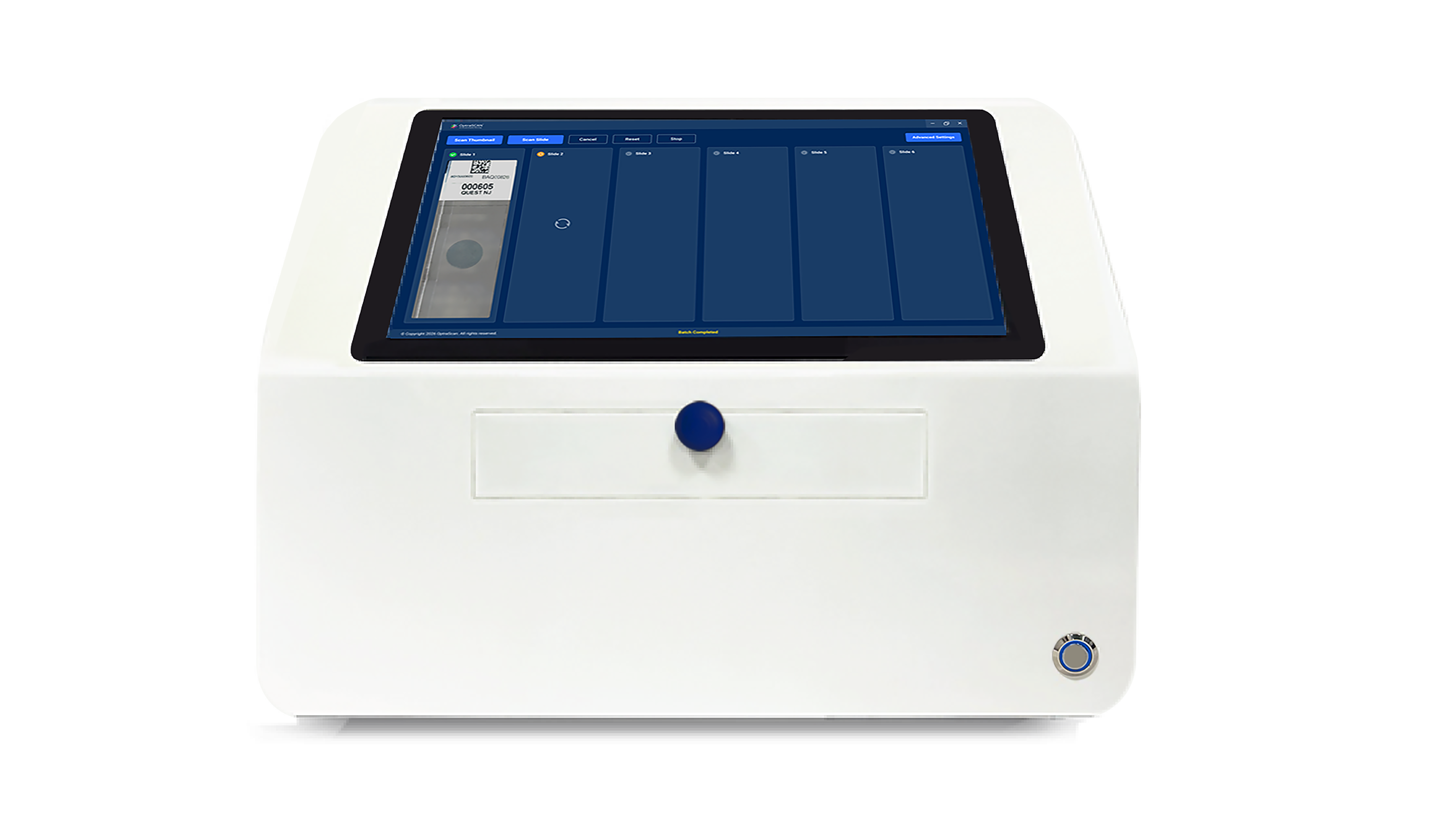
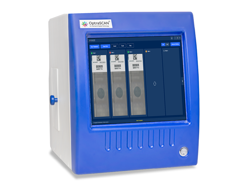
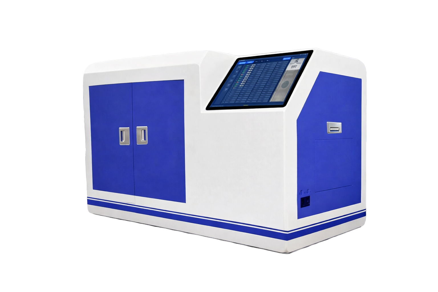
Works with OS-SiX (6 slides) through OS-Ultra (480 slides). Z-stacking, AI autofocus, and composite imaging built in.
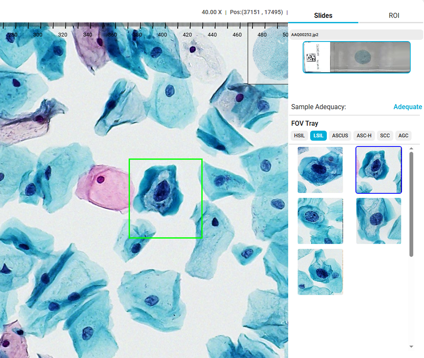
Flags normal cases, identifies HSIL, LSIL, ASCUS, ASC-H, SCC, and AGC — ranking atypical cells by probability for focused review.
Clinician-ready reports with thumbnails, classification stats, and flagged regions. Full EMR/LIS integration available.
Mobile-ready and cloud-connected. From national labs to mobile screening units in underserved communities.
A seamless digital workflow from prepared slide to clinician-ready report — powered by OS-SiA™, the world's first patented technology that scans, indexes, and analyzes simultaneously.
CytoSiA's AI engine classifies individual cervical cells according to the Bethesda system, ranking each by probability of abnormality. Pathologists see the most critical findings first — reducing review time, fatigue, and missed detections.
Reports are generated automatically with visual summaries, classification statistics, and flagged regions — ready for clinical review or EMR/LIS integration.
Unlike traditional digital cytology tools, CytoSiA is designed from the ground up for scalability, accessibility, and real-world deployment — from national programs to mobile outreach clinics.
Flags high-confidence normal cases automatically, freeing pathologists to focus on atypical and borderline results.
Atypical cells ranked by AI confidence — the most suspicious findings surface first for immediate attention.
Optimized for OptraSCAN scanners but customizable for third-party systems and existing digital images.
Extend screening into underserved communities with cloud-connected, telepathology-enabled mobile deployment.
Deep learning models trained on millions of cervical cancer datasets for high sensitivity and specificity.
Structured, clinician-ready reports with Bethesda classification, visual summaries, and EMR/LIS integration.
The WHO has set ambitious 90-70-90 elimination targets by 2030. CytoSiA helps screening programs close the gap between where they are and where they need to be.
Purpose-built for the settings driving cervical cancer elimination — from national screening infrastructure to last-mile mobile units.
Scale cervical cancer screening to reach millions of women with consistent, standardized AI-assisted triage and automated reporting across the entire program.
Fast, cost-effective triage for low- and middle-income countries where trained cytotechnologists are scarce and screening demand is highest.
Mid- to high-volume labs improving throughput and consistency. Reduce reader fatigue and missed detections while maintaining clinical quality.
Telepathology-ready mobile deployment extending screening into underserved communities. Cloud-connected for remote expert review and collaboration.
See how AI-powered digital cytology can transform your cervical cancer screening program — from slide to structured report in minutes, at any scale.