OS-FS is a frozen section imaging modality by OptraSCAN®, purpose-built for real-time intraoperative pathology review. Delivered through compatible OptraSCAN platforms such as OS-SiX™ and OS-Lite™ , OS-FS enables live digital microscopy using Live View Mode as slides are prepared. With a compact footprint suitable for surgical suite environments, OS-FS preserves established diagnostic* practices while extending immediate local and remote access to frozen section review, without relying on archived whole slide imaging.
Frozen section workflows demand immediate access to expert interpretation, seamless collaboration, and minimal disruption to surgical timelines. Traditional approaches rely on physical microscopes and on-site expertise, limiting flexibility and access, particularly across hospital networks and distributed care environments.
OS-FS supports pathologist-led interpretation in speed-critical environments, eliminating any additional unnecessary scanning, storage, or workflow complexity.

Frozen section slides are prepared using standard intraoperative protocols.

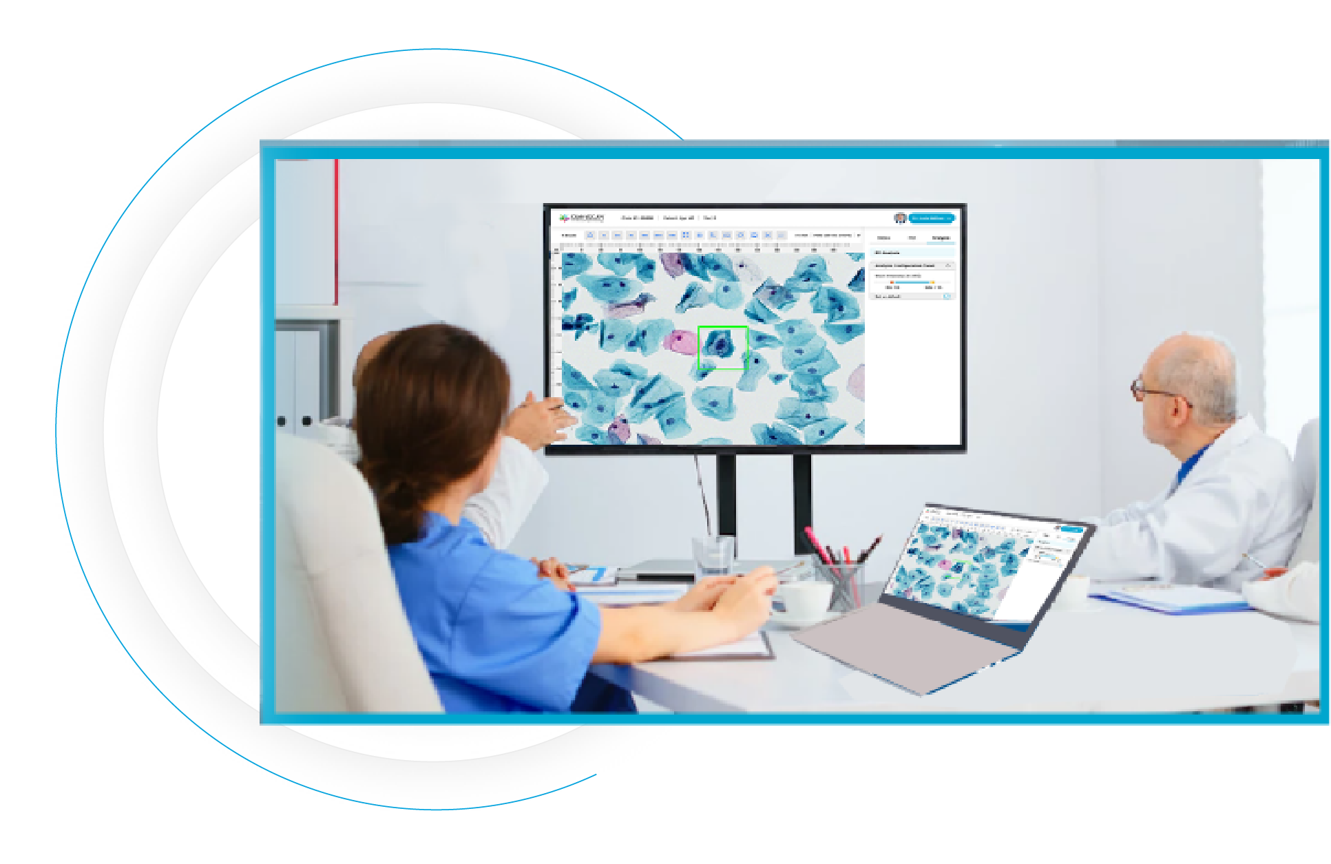
Slides are visualized instantly using Live View Mode for real-time digital microscopy.

Pathologists can review and consult remotely across locations.

Findings are communicated immediately to support surgical decision-making.
Real-time slide visualization using Live View Mode without post-processing delays
Optimized for surgical workflows requiring immediate access and responsiveness without any complexity
Enables real-time consultation and collaboration across hospital networks
Preserves established diagnostic practices with full control over review and reporting
Automated focus setting and tissue detection support efficient live visualization
AI-powered detection enhances visualization accuracy and consistency
Patented composite imaging and Z-stacking capabilities improve visualization of uneven specimens
Supports 1” × 3”, 2” × 3”, 3” × 3”, and 4” × 3” slide formats
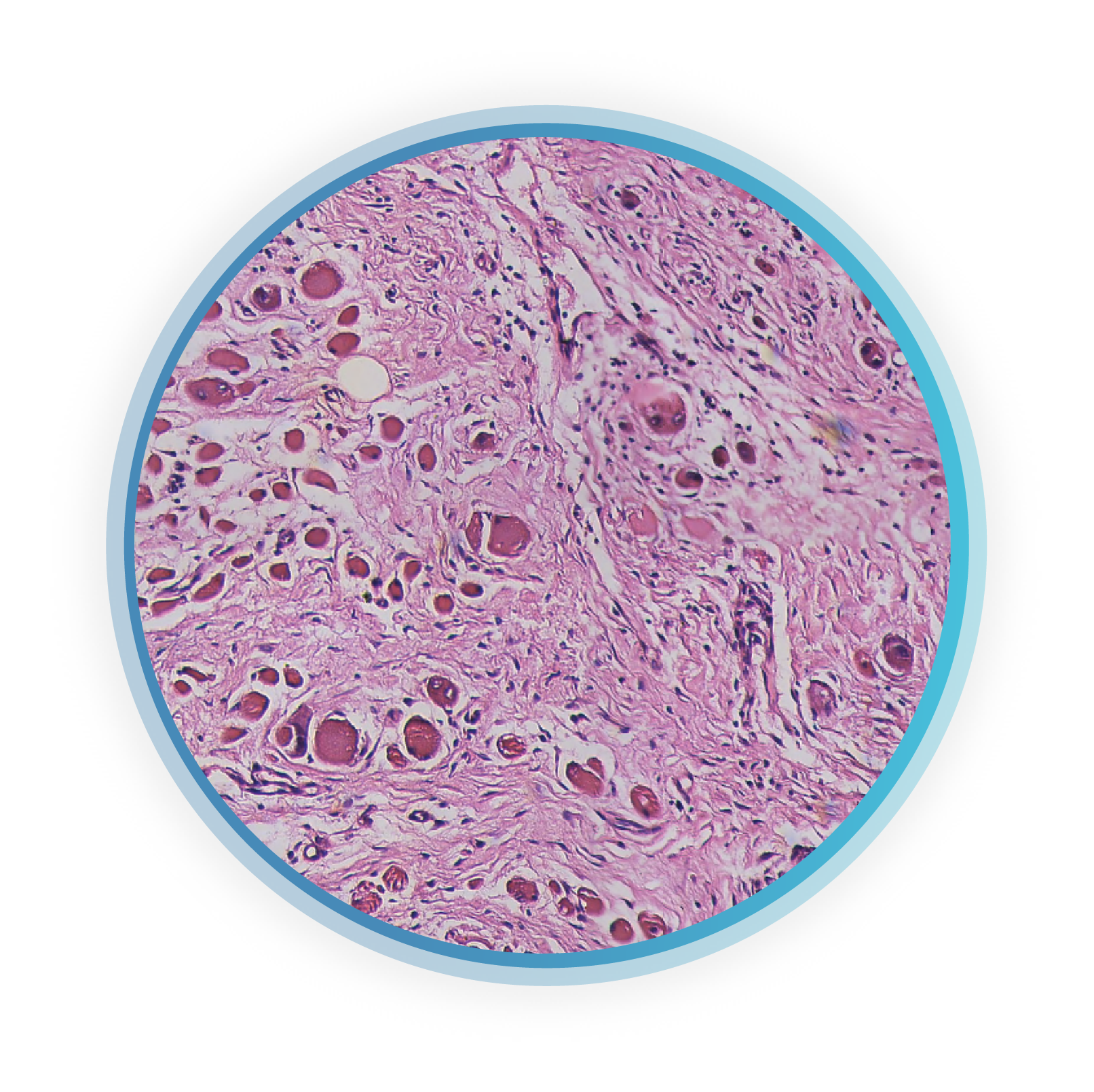
Example of a frozen section specimen visualized in real time using OS-FS Live View Mode, enabling immediate digital review during intraoperative workflows
OS-FS is available as a frozen section imaging modality on OptraSCAN's compact footprint scanners suitable for surgical settings:
When enabled, OS-FS integrates seamlessly with OptraSCAN's digital pathology ecosystem:
OS-FS supports pathologist-led interpretation in speed-critical environments, eliminating any additional unnecessary scanning, storage, or workflow complexity.

Supports frozen section consultation and rapid cytology/FNA review

Surgical pathology workflows requiring immediate expert feedback

Extends expert access without changing established pathology practices

Enables connected intraoperative workflows without operational disruption.
Please fill out the form below and complete all questions with an asterisk (*)


