An automated image analysis algorithm that evaluates PD-L1 expression on both tumor and immune cells — bringing objectivity, consistency, and efficiency to lung cancer IHC research.
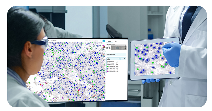
OptraSCAN's PD-L1 Lung Cancer Algorithm is an automated image analysis tool that evaluates PD-L1 expression by detecting both partial and complete membrane staining on tumor cells, as well as membrane and cytoplasmic staining on immune cells — all within a single whole-slide scan.
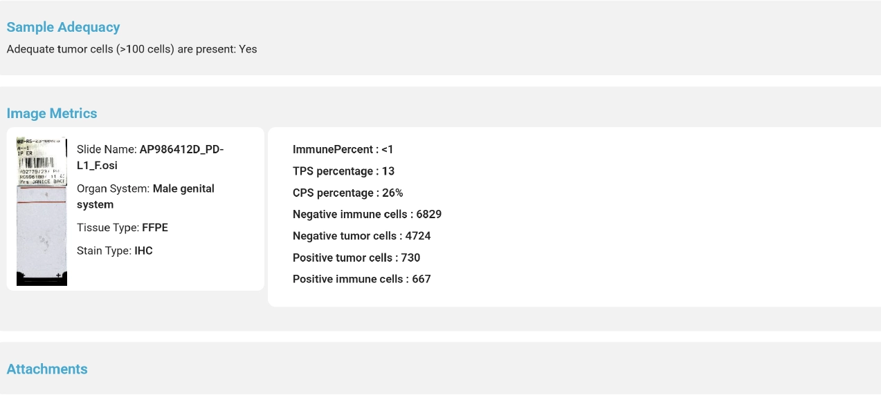
The algorithm calculates PD-L1-positive cells (tumor, lymphocytes, and macrophages) relative to total tumor cells, delivering a comprehensive view of PD-L1 expression across the entire tissue section.
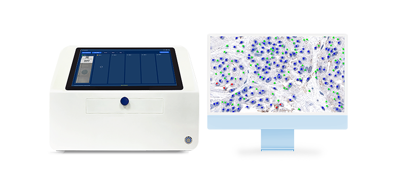
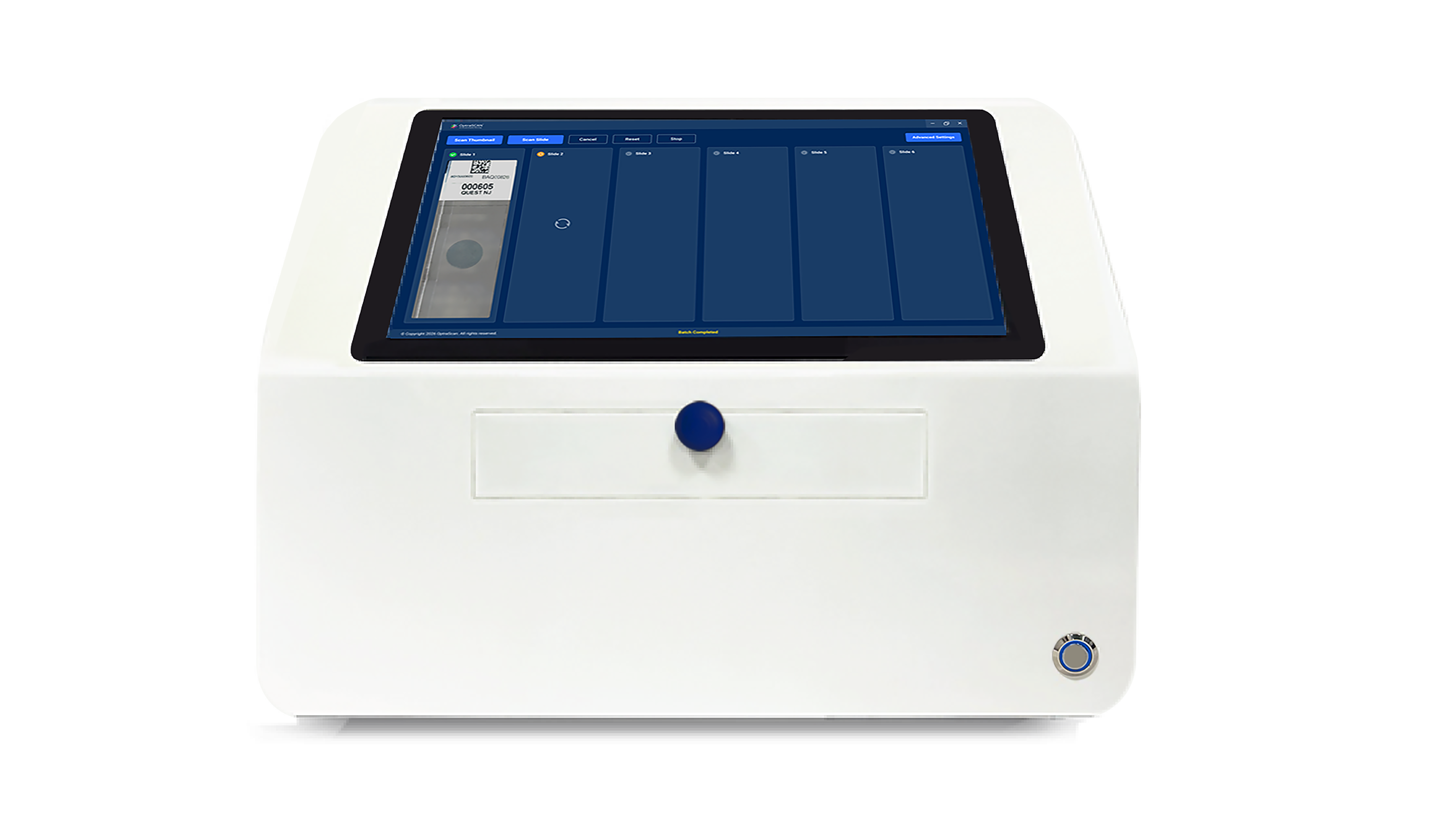
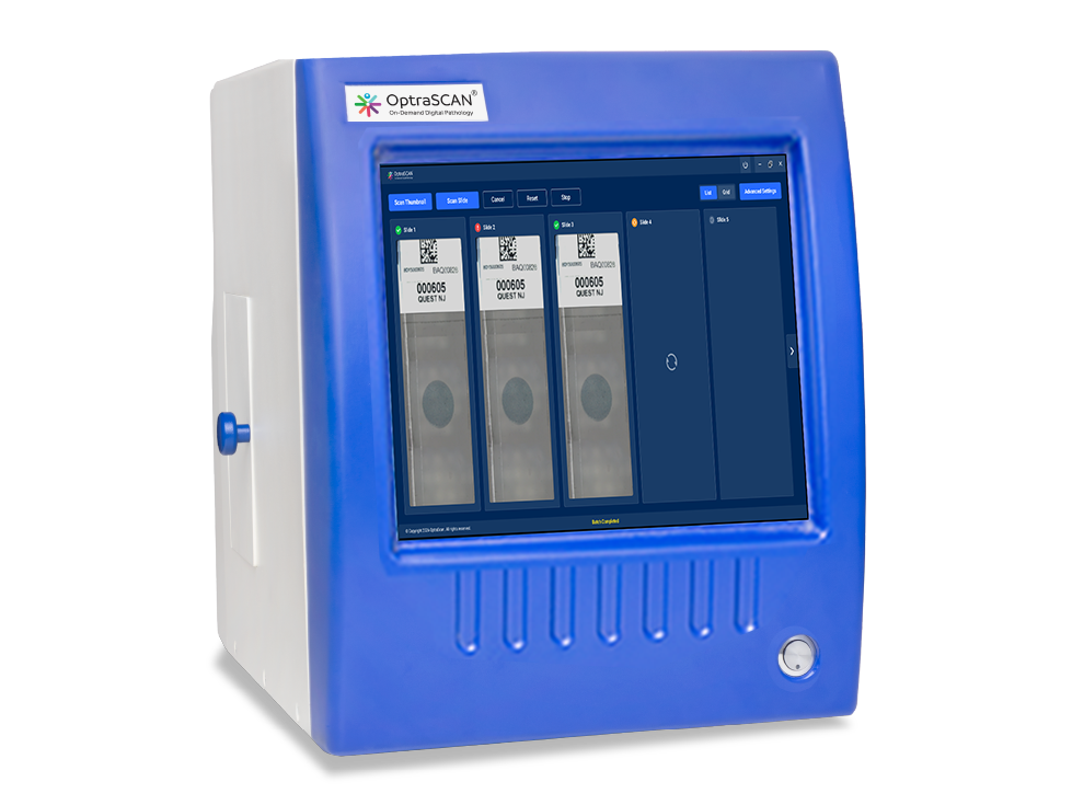
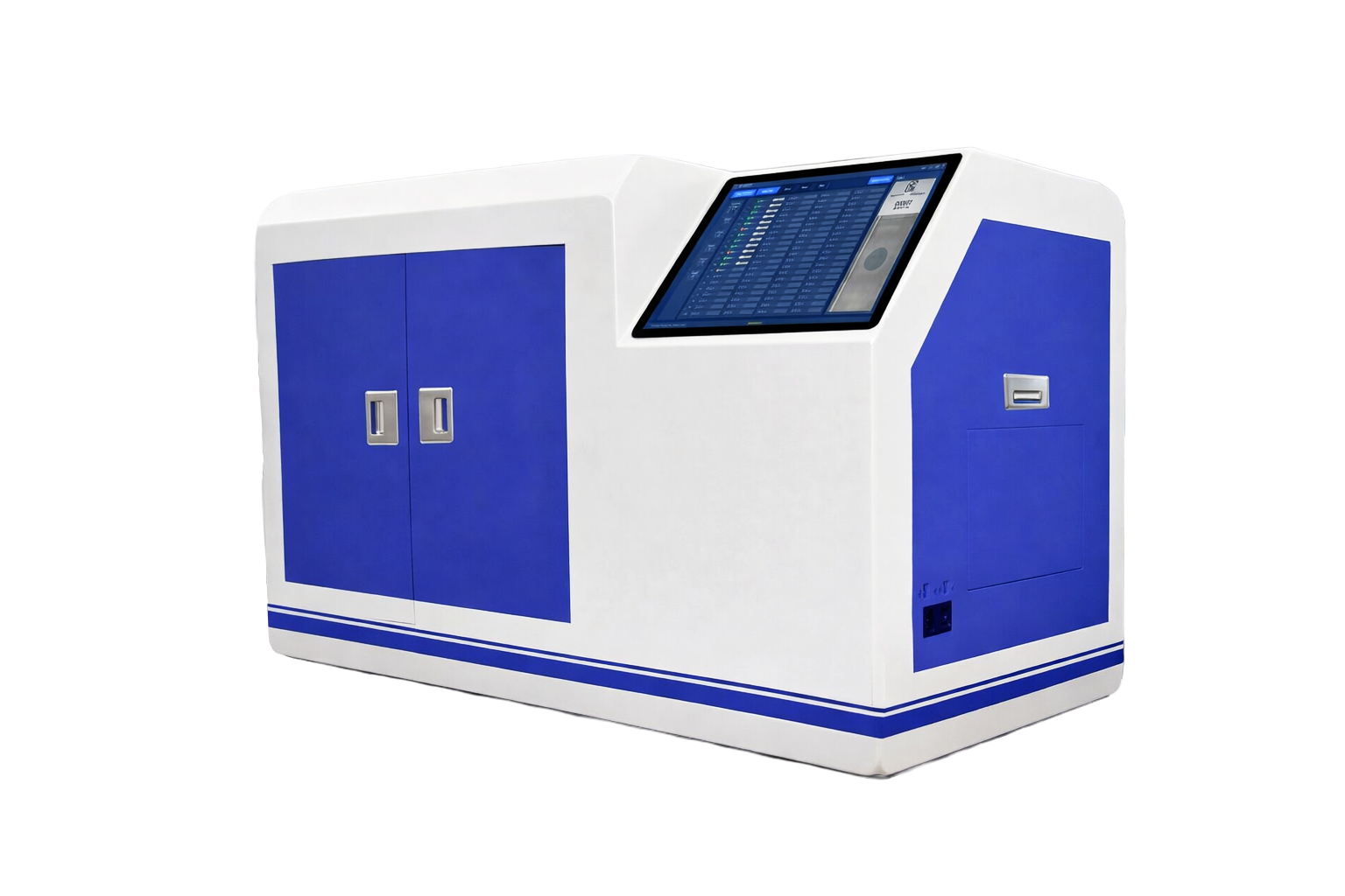
High-resolution whole slide images captured using OptraSCAN scanners, preserving cellular detail at 20× or 40×.
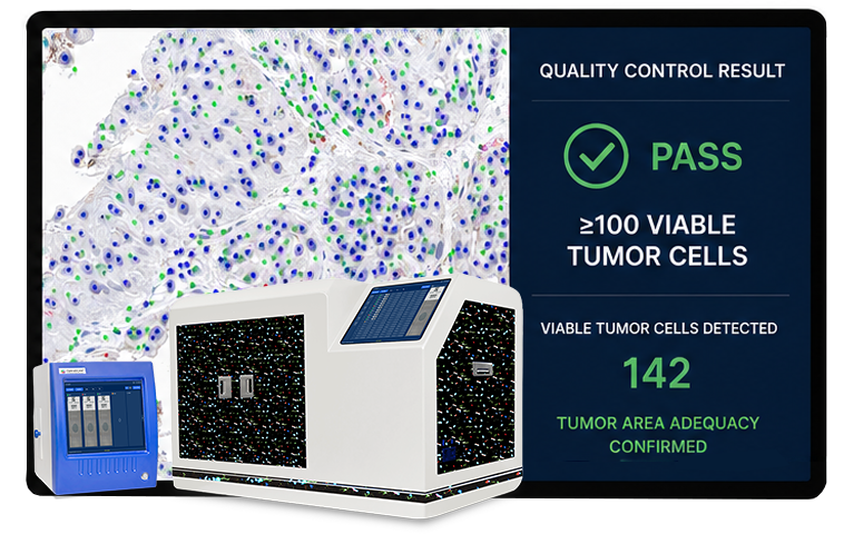
Automated validation confirms ≥100 viable tumor cells using a neural-network-powered region detection system.
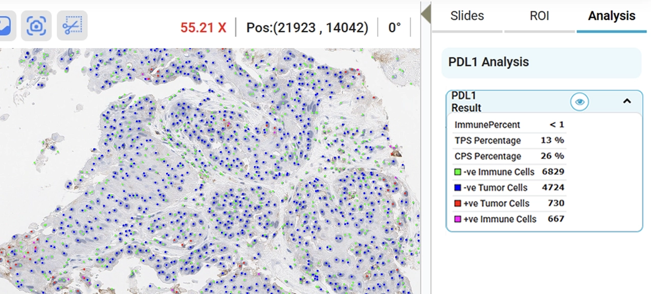
PD-L1 expression patterns assessed across membrane and cytoplasmic staining. Positive cells categorized by type.
Detailed scores and structured reports generated automatically for documentation and downstream analysis.
The PD-L1 algorithm evaluates positive cells on both tumor and immune cells with minimal human intervention — generating reproducible results that support objective, standardized analysis across samples and studies.

Calculates PD-L1-positive tumor cells, lymphocytes, and macrophages relative to total tumor cells — delivering a complete view of PD-L1 expression across tumor and immune cell populations in a single analysis.
Integrated with OptraSCAN's brightfield and fluorescence scanners, the algorithm assesses tumor area adequacy by confirming ≥100 viable tumor cells — leveraging a computer-aided region detection system powered by an artificial neural network.
A case management and reporting module streamlines documentation and results handling — enabling efficient data management across research workflows with secure, structured outputs ready for publication or further analysis.
Automated analysis minimizes subjective variation, supporting more consistent PD-L1 assessments across readers, sites, and studies.
Automated evaluation frees researchers to focus on interpretation and discovery — reducing turnaround time without sacrificing quality.
Accurate, reproducible PD-L1 expression data supports effective research decisions and facilitates meaningful advances in lung cancer understanding.
OptraSCAN's PD-L1 Lung Cancer Algorithm is built on rigorous research and validated to ensure the highest standards of accuracy and reliability. Our work, published in the prestigious Nature journal, underscores the technology's significance in advancing lung cancer research.
Through continuous refinement and partnerships with top institutions and clinical experts, the algorithm stays aligned with the latest scientific breakthroughs — providing researchers with cutting-edge tools for precision PD-L1 analysis.

See how automated PD-L1 expression analysis can bring objectivity and efficiency to your IHC research workflow.
Request a Demo