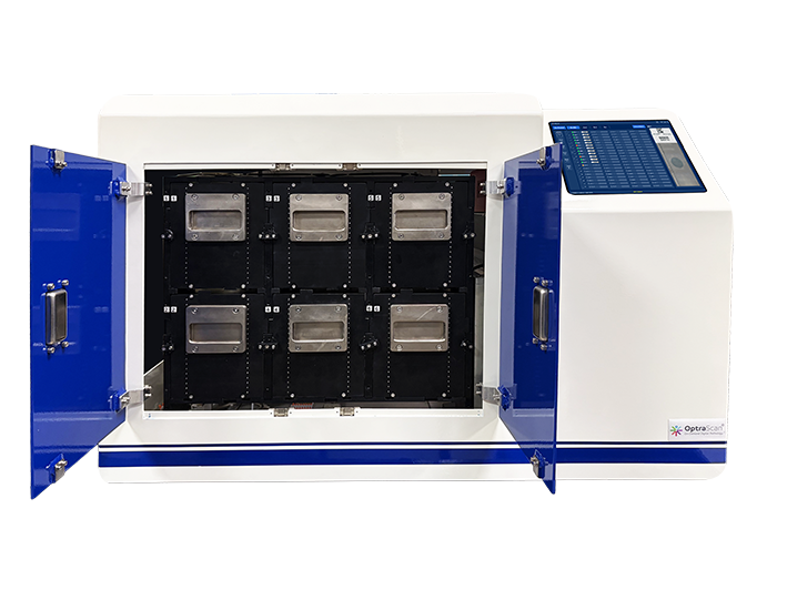
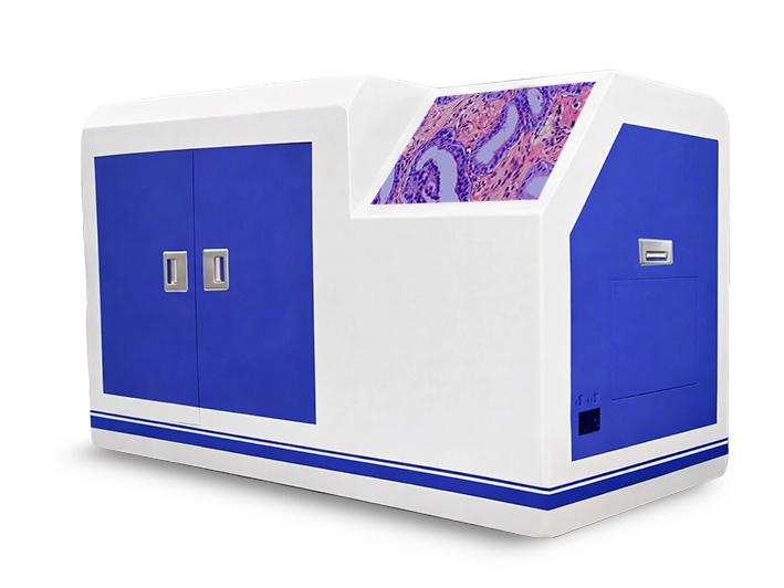
The fastest scanner in the OptraSCAN lineup. Scan up to 480 slides with walk-away automation, no-touch loading, and built-in AI — delivering whole slide images in under 60 seconds at 40×.
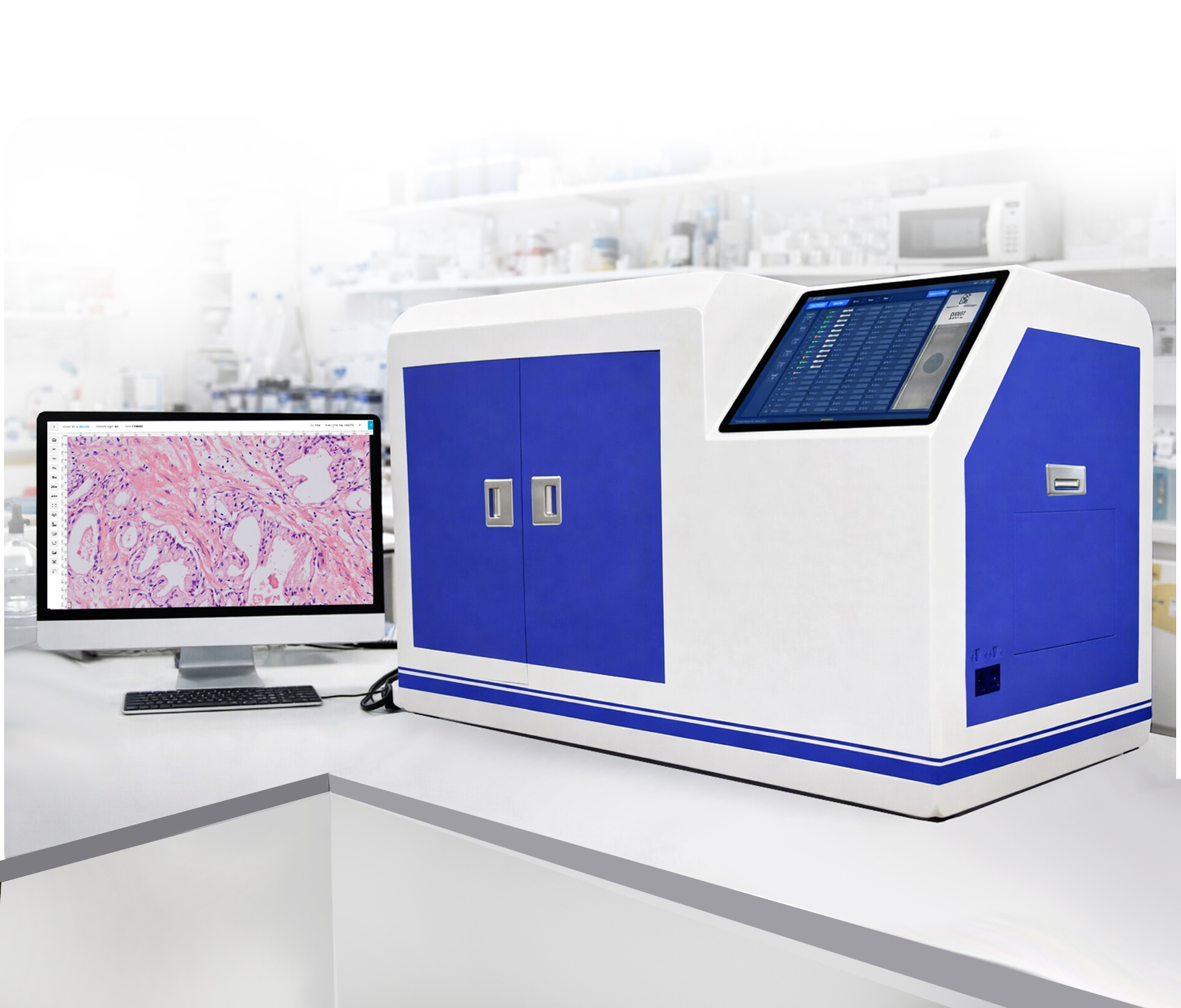
The OS-Ultra serves research, clinical, educational, and telepathology workflows — all from a single platform with enterprise-grade throughput.
High-resolution imaging of tissue samples for in-depth studies. Capture cellular structures and morphology with exceptional clarity.
Build vast digital slide archives for learning, training, and mentoring. Enable collaboration and remote supervision across institutions.
Remote access and interpretation via TELEPath. Share cases, discuss findings, and seek second opinions without physical transport.
Leverage AI-powered algorithms for accurate quantification. Eliminate inter-reader variability for consistent, precise results.
Cloud or local storage for long-term archival. Eliminate physical slide maintenance while ensuring instant accessibility.
Walk-away automation for 80–480 slides. No-touch loading reduces errors and contamination. Designed for 24/7 operation.
| Specification | Detail |
|---|---|
| Imaging Mode | Brightfield |
| Slide Capacity | 80 / 160 / 240 / 320 / 400 / 480 configurations |
| Scan Time (15×15mm) | 30s at 20× | 60s at 40× |
| Slide Formats | 1×3″, 2×3″, 3×3″, 4×3″ |
| Magnification | 20×, 40× |
| Image Output | JPEG2000, TIFF, oTIFF, DICOM |
| Weight | 233.6 lbs (106 kg) |
| Dimensions | W: 24.2″, L: 37.4″, H: 24.8″ |
| Tissue Samples | Cytology, histology, hematology, special stains, sputum |
| AI / Software | Fully compatible with OptraSCAN IMS & AI analytics |
See how the fastest scanner in our lineup can transform your high-volume pathology workflow.
Request a Demo