
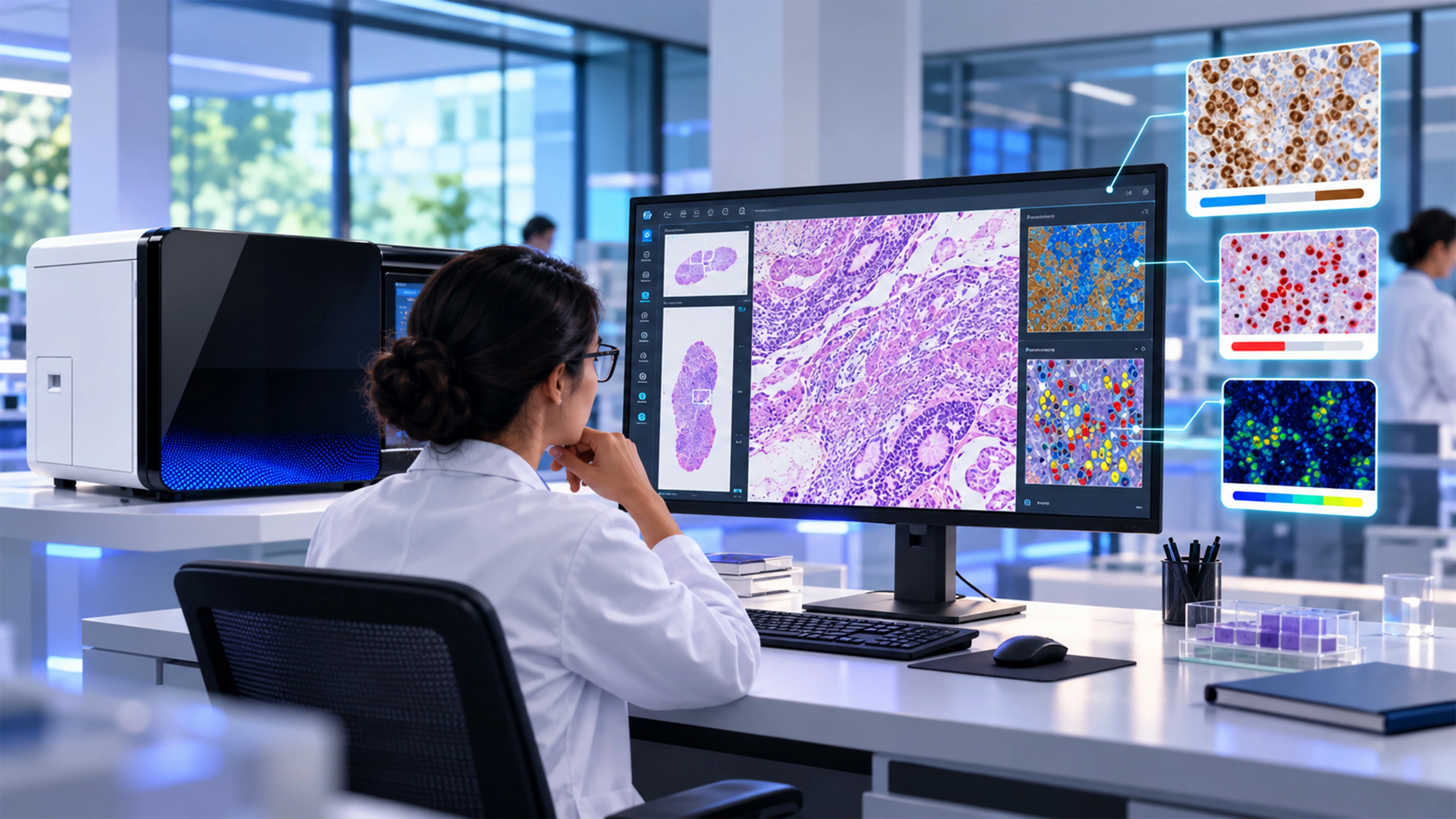
If you're a clinician outside of pathology — an oncologist, a surgeon, a hospitalist — "computational pathology" is one of those terms that shows up in a tumor board slide or a vendor pitch, gets nodded along to, and rarely gets actually explained. That's a shame, because the underlying idea is simpler than the name suggests, and understanding it makes a real difference in how you interpret the reports and biomarker scores landing on your desk. Here's the plain-language version.
Strip away the jargon, and computational pathology is this: taking a digitized image of a tissue sample, and having a computer analyze it alongside other patient data — clinical history, lab results, sometimes radiology or molecular data — to help generate a more accurate, more consistent diagnostic or prognostic answer than a human working from the slide alone would produce as quickly.
It's not one single technology. It's better understood as three layers stacked on top of each other:
That third layer is what separates computational pathology from just "a scanner took a picture." It's the step where machine learning in pathology actually earns its keep — not by replacing a pathologist's read, but by handling the parts of image analysis that are repetitive, high-volume, or prone to natural human variability.
Machine learning in pathology shows up specifically in that second layer — the image analysis step. Deep learning models, trained on large sets of labeled tissue images, learn to recognize patterns: tumor cells versus normal tissue, mitotic figures, staining intensity for a specific biomarker. Because these models don't get tired, distracted, or inconsistent from one slide to the next the way any human evaluator naturally can, they're particularly well suited to quantitative, repeatable scoring tasks — the kind that benefit from being done the exact same way every single time.
This is also where AI in pathology tends to get misunderstood. The models aren't making an independent diagnosis — they're producing a structured, quantified output (a cell count, a percentage of positive staining, a flagged region of interest) that a pathologist then reviews, interprets in clinical context, and signs out. The AI narrows and standardizes the input to the pathologist's judgment; it doesn't substitute for it.
For a clinician receiving a pathology report, understanding this layered structure changes how you read what lands in front of you. A biomarker score generated with computational assistance — say, a quantified HER2 or Ki-67 result — should read as more standardized and more reproducible across cases than a purely manual visual estimate, because the scoring criteria were applied identically every time rather than varying slightly by reviewer or by day. That consistency is directly relevant to treatment decisions that hinge on a specific threshold or cutoff score.
In practice, computational pathology only works if the digitization, image analysis, and reporting pieces are actually connected — not separate tools bolted together after the fact. OptraSCAN's platform is structured around exactly that chain: high-throughput scanners like OS-Ultra handle the digitization step, IMAGEPath manages and stores the resulting whole slide images, and OptraSCAN's AI-enabled image analysis tools — including validated biomarker scoring for HER2, ER, PR, Ki-67, and other markers — handle the quantitative layer, producing standardized results a pathologist reviews before anything reaches a report. The platform is built AI-ready end to end, so a lab isn't stitching together a scanner from one vendor and a separate analysis tool from another to get a computational pathology workflow that actually functions clinically.
Computational pathology isn't a mysterious black box — it's digitization, targeted image analysis, and data integration, applied in a way that makes quantitative pathology results more consistent than manual estimation alone. Understanding that layered structure is really all you need to interpret what a computationally-assisted result is actually telling you, and where a pathologist's judgment still sits on top of it.
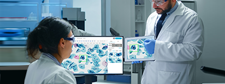
Improving diagnostic outcomes through digital workflow adoption.

Everything you need to know about digitizing histopathology workflows.
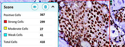
Where AI stands today and what's needed to close the gap.