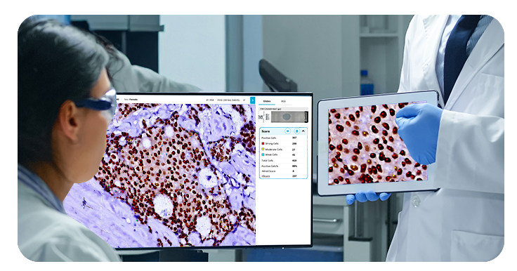
Scan, analyze, and auto-segregate breast cancer IHC cases in a single workflow. Multi-biomarker scoring with automated ASCO-CAP categorization — powered by OS-SiA™ technology.
HistoSiA Breast combines whole-slide imaging with real-time AI to scan, analyze biomarker expression, and auto-segregate IHC cases into diagnostic* categories — before the pathologist even begins review. A scalable, multi-biomarker platform for HER2, ER, PR, Ki-67, AE1/3, EGFR, and p53.

OS-SiA™ technology performs IHC quantitative analysis across entire tissue sections during the scanning process itself.

AI classifies HER2 slides into 0, 1+, 2+, and 3+ categories during scanning — enabling immediate case triage.

Scalable platform integrating HER2, ER, PR, Ki-67, AE1/3, EGFR, and p53 analyses within the same digital workflow.

Flexible pricing — pay only for slides analyzed. Minimum upfront investment, maximum operational flexibility.
Powered by OS-SiA™ — scan, index, and analyze IHC slides simultaneously in a single pass.
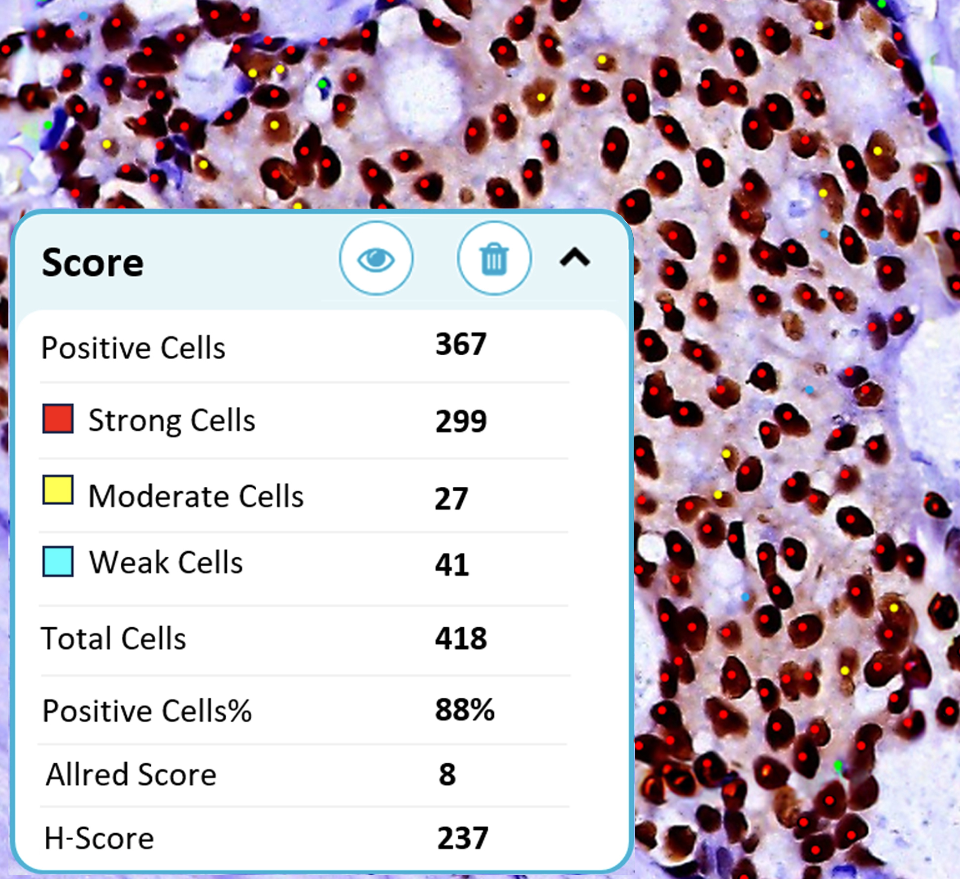
HistoSiA delivers interpretable analytical results for real-world pathology workflows — algorithm overlays highlighting biomarker expression, numerical scoring outputs, and whole-slide analysis across representative tissue regions.
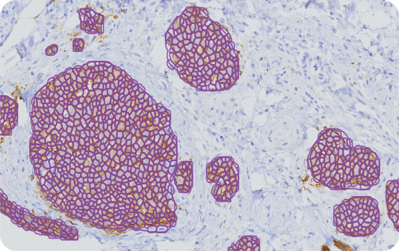
Automated categorization into 0, 1+, 2+, and 3+ with ASCO-CAP-aligned scoring and visual membrane overlays.
ASCO-CAP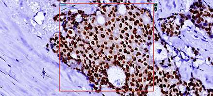
Quantitative nuclear staining analysis with Allred and H-Score assessment across tissue sections.
Allred · H-Score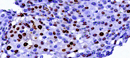
Hormone receptor quantification with standardized scoring aligned to clinical interpretation guidelines.
Allred · H-Score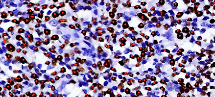
Proliferation index assessment with automated cell counting and percentage quantification across hotspot regions.
Proliferation IndexHistoSiA integrates established clinical scoring methodologies directly into the analysis workflow — Allred, H-Score, ASCO-CAP, proliferation index, and TNM staging. All results are managed digitally within IMAGEPath® with visual overlays and exportable reports.
Supports quantitative and semi-quantitative IHC analysis aligned with CPT® 88361 coding for per-specimen, per-antibody interpretation. Coverage and reimbursement vary by payer and institution.
HistoSiA's scalable architecture supports quantitative pathology across multiple disease areas. Labs can add new biomarkers and analyses without reinvesting in new hardware or workflows.
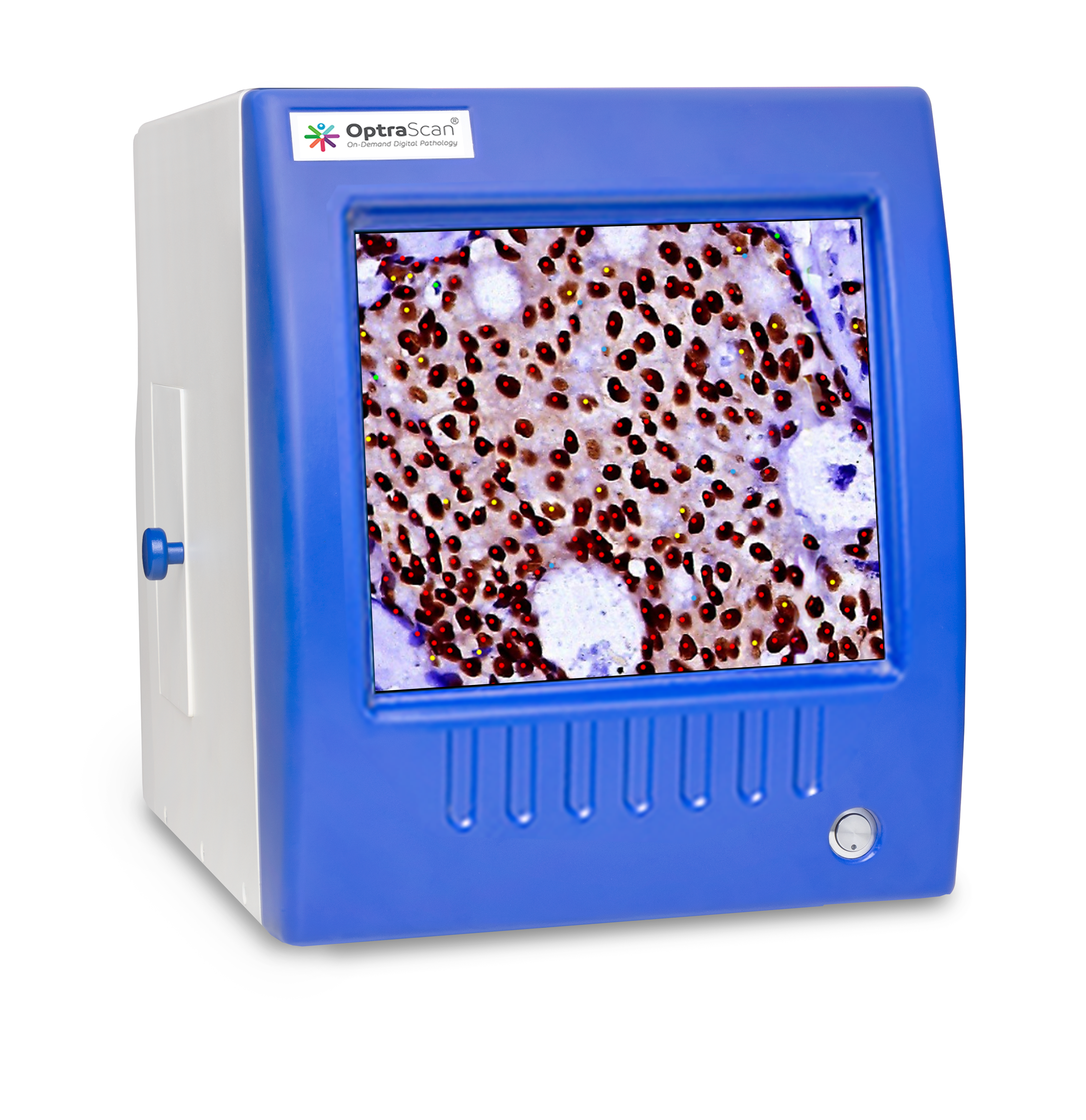
Start with HER2 and expand to ER, PR, Ki-67, AE1/3, EGFR, and p53 as your lab's needs evolve — all within the same platform.
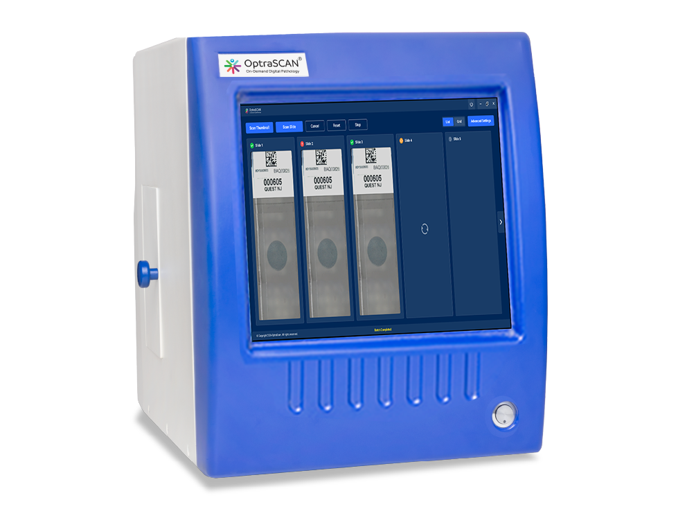
Optimized for OptraSCAN systems but designed to integrate with existing digital pathology infrastructure. No vendor lock-in.
All biomarker analyses, scoring, and case management in one environment. Eliminates fragmented tools and manual interpretation variability.
Supports quantitative IHC workflows aligned with established coding. CPT® 88361 per-specimen, per-antibody. Coverage varies by institution.
Purpose-built for pathology teams working with breast cancer biomarkers — from early digital adopters to high-volume clinical labs.

Labs beginning to integrate AI-assisted analysis for breast cancer biomarker interpretation — with a low-risk, pay-per-use entry point.

Labs with current or planned digital pathology infrastructure looking to add automated IHC quantification and auto-segregation.

Teams needing reproducible, consistent biomarker scoring across multiple readers, sites, and workflows for clinical or research applications.

Academic institutions and translational research groups studying breast cancer biomarkers with quantitative, digitally tracked outputs.
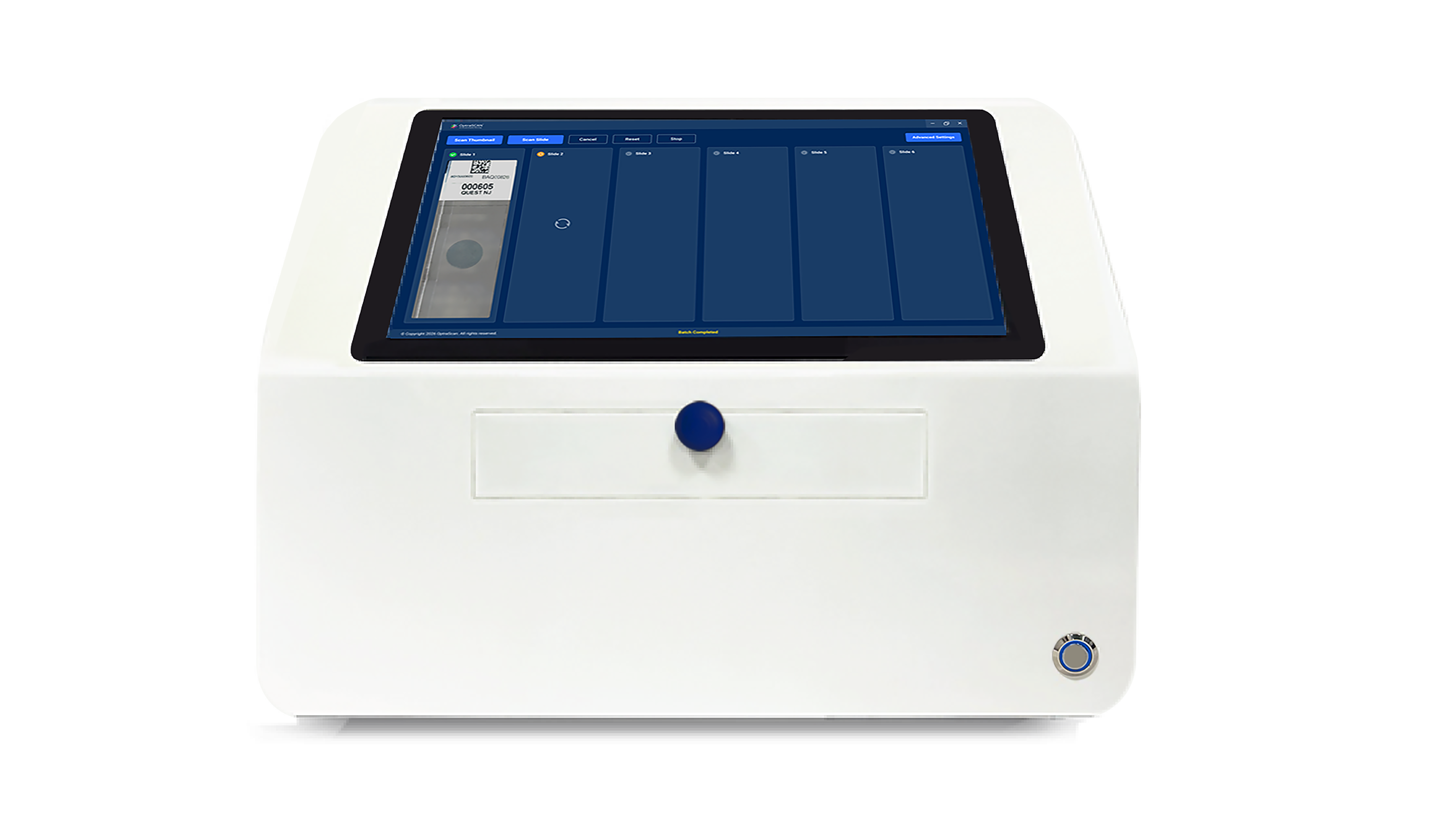
See how automated biomarker scoring and auto-segregation can streamline your breast cancer pathology workflow — pay only for what you use.