
In recent years, digital pathology has emerged as a pivotal technology reshaping healthcare globally. Leveraging advanced imaging and artificial intelligence (AI), digital pathology enhances diagnostic accuracy, efficiency, and collaboration among healthcare professionals and researchers.
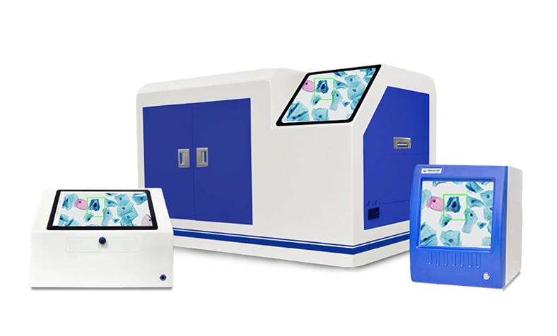
Pathology, the field that has brought us so many medical breakthroughs, has always been at the forefront of human discovery. The countless health challenges we've overcome thanks to this knowledge and technology are impressive. One of the latest advancements shaking up the scene is digital pathology, which has radically redefined how we diagnose and address survival concerns with incredible precision.
Digital pathology has completely transformed the practice. With the emergence of digital slide scanners, tasks like:
Digital pathology streamlines specimen acquisition and digitization for improved accessibility and workflow management.
Digital systems simplify pathology data and slide management through centralized storage and retrieval.
Pathologists can easily share digital slides for consultations and collaborative diagnostics across locations.
AI-assisted analysis improves interpretation accuracy and supports better diagnostic decisions.
This is a big shift from the old-school analog methods, where pathologists relied on physical glass slides and manual microscope-based examinations.
In the old days, pathologists would peer through a microscope at tissue samples, make their observations, and jot down findings on paper or into a computer. This process was slow, labor-intensive, and error-prone. Especially, those physical slides were at risk of being damaged, lost, or misplaced, causing delays in diagnosis and treatment.
But digital pathology uses computer-based technology to tackle these issues head-on. With virtual microscopy, pathologists can now view and analyze tissue samples on a computer screen instead of through a traditional microscope. This modern approach offers significant benefits like improved accuracy, efficiency, and accessibility.
Digital pathology scanners are automated, Whole slide imaging scanner systems designed to capture high resolution images (WSIs) of histology and cytology samples prepared on glass slides.
Digital pathology slide scanner employ a sophisticated array of high-powered optics, specialized illumination, and precision stage movement to capture whole-slide images (WSIs) at magnifications that surpass conventional microscopy. These digital renderings preserve the entirety of the tissue section on the pathology slide.
Traditional pathology relies on the direct microscopic examination of glass slides by pathologists. This method, while highly effective, presents certain limitations.
Pathology slide scanners are like high-tech assembly lines for pathology. They combine several powerful tools specifically designed to analyze tissue samples and create top-notch digital versions.
These include intense-grade lenses and objectives that are designed to capture whole slide images of the tissue samples at various magnifications.
The motorized stage is responsible for moving the glass slide automatically during the scanning process.
The digital pathology scanner's beating heart is a high-resolution camera that captures magnified snapshots of the tissue.
The software manages the scanning process, processes captured images, stitches them together, and stores the resulting digital slide.
Placing the glass slide on the motorized stage of the digital pathology scanner.
Selecting the desired magnification for scanning.
Initiating the scanning process depending on the pathology scanner model.
The motorized stage moves automatically while the digital camera captures overlapping images.
The software stitches together individual images into a single digital slide.
The resulting slide is stored securely and can be viewed, shared, and analyzed digitally.
Whole slide imaging enables detailed zoom capabilities, improving the identification of subtle morphological features and biomarker expression patterns.
Whole slide imaging scanners enable remote access, collaboration, consultations, and second opinions from specialists worldwide.
Digital pathology reduces physical storage requirements while improving workflow speed and laboratory productivity.
Advanced image analysis software enables objective measurement of tissue features and biomarker expression for research and personalized medicine.
Digital slides scanner can be used for primary diagnosis, second opinions, and remote consultations.
Pathology slides can be used for research, training, and teaching purposes.
Digital pathology supports efficient analysis of tissue samples for new drug development and clinical trials.
Digital slides can be used for remote diagnosis and consultations for underserved and remote areas.
The emergence of an increased number of digital pathology solutions represents a significant shift in tissue analysis. With its high-end features and smart computing qualities, a digital pathology slide scanner enables collaboration among geographically dispersed pathologists.
OptraSCAN is a pioneering digital pathology company that paid its dues to pathological and research professionals and will continue to do so with the same dedication. To avail yourself of our feature-rich digital pathology slide scanner, contact us today and help your laboratory provide the best diagnostic* results to your patients.
Contact Us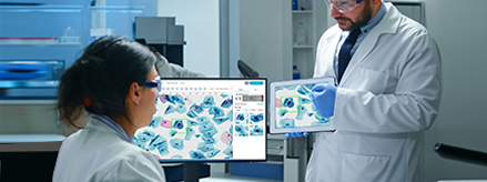
Improving diagnostic outcomes through digital workflow adoption.

Everything you need to know about digitizing histopathology workflows.
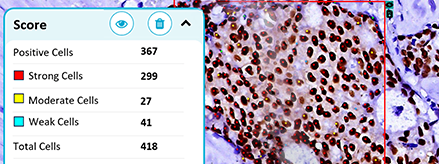
Where AI stands today and what's needed to close the gap.