The term "digital histopathology" describes the application of digital technology to pathology, particularly for tissue sample examination and analysis. In traditional histology, a pathologist examines tissue samples under a microscope in order to make diagnoses*, assess the degree to which a disease has progressed, and inform treatment choices.
The technique of digital histopathology is improved and made easier by the digitization of the histological slides. High-resolution histology slide scanner is used to digitally scan the images of the tissue samples on glass slides rather than using a microscope to observe them. After that, these digital photos can be electronically shared, analyzed, and archived.
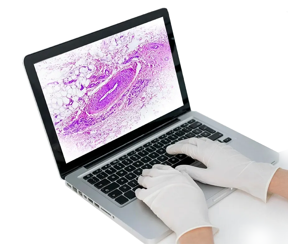

Top-notch histology scanner makes tiny pictures of whole histology slides at different magnifications.

Digital photos are easily retrieved and archived because they are kept in a digital database. This makes it easier for pathologists to collaborate and view the photos remotely.

Thanks to digital pathology, pathologists can use software tools and algorithms for image analysis to help with quantitative analysis, pattern recognition, and other sophisticated studies. This can help with more repeatable and objective assessments.

Experts from different places can collaborate and conduct telepathology consultations, second opinions, and collaborative work because pathologists can view and interpret digital slides remotely.

Digital histopathology is helpful for teaching medical professionals. Students and trainees can study a wide range of situations by exchanging digital slides for educational reasons.
Among the many benefits of digital histopathology are increased accessibility, better teamwork, and the possibility of more effective and standardized diagnostic* procedures. Ongoing development and research in this sector are still needed to address issues like the need for validated digital tools, standardizing digital pathology methods, and data storage requirements.
While digital pathology offers many benefits, solving issues like standardization, data security, and tool validation is critical to ensuring its widespread and successful adoption in clinical practice.
Histopathology, which entails examining tissues and cells under a microscope, offers essential insights into the causes of various diseases. The following are some of histopathology's main benefits:
Histopathology is a key component of medical diagnosis*. It enables precise identification and description of a wide range of illnesses, including infections and cancers.
Clinicians use histopathological findings as guidance when they plan appropriate therapies, such as radiation therapy, chemotherapy, surgery, or other procedures.
Staging is useful in determining the severity and stage of illnesses, especially cancer. It guides prognostic information and treatment decisions.
Tumor classification, cellular origin identification, and malignancy classification depend on histopathology.
Histopathological findings can provide prognostic information, helping to forecast the probable course of a disease and the patient's final outcome.
Histopathology-observed changes in tissue morphology can help guide therapeutic approach modifications by providing insight into how effectively a patient responds to treatment.
In biomedical research, histopathology plays a crucial role in understanding disease causes and developing new therapeutic approaches.
Histopathological slides facilitate the study of normal and aberrant tissue structures and are vital educational tools for medical students, residents, and other healthcare workers.
Histopathology is a confirmatory tool that adds to a thorough understanding of a patient's condition by confirming or elaborating on clinical suspicions.
By offering insights into individual differences in disease presentation, histopathology promotes personalized medicine by enabling customized treatment regimens.
It plays a crucial role in identifying and detecting infectious pathogens and supporting the diagnosis* of various contagious disorders.
Histopathology is used in forensic medicine to identify injuries, ascertain the cause of death, and give vital information for legal investigations.
Histopathological brightfield slide scanner archives, both physical and digital, provide long-term preservation and support quality control, follow-up evaluations, and retrospective research.
Although histology provides many benefits, it's vital to remember that it also has drawbacks. It is frequently supplemented by additional diagnostic* techniques like imaging investigations, clinical assessments, and molecular tests to provide a thorough picture of a patient's condition.
The histology scanner smoothly integrates the real-time analysis of cell samples and scanning, which is a revolutionary feature.
Discover the pathology of the future with OptraSCAN's state-of-the-art AI technologies, which will transform the precision and effectiveness of diagnosis*. We provide solutions intended to improve productivity and efficiency by streamlining and optimizing your lab workflow. Contact us to revolutionize pathology workflows.