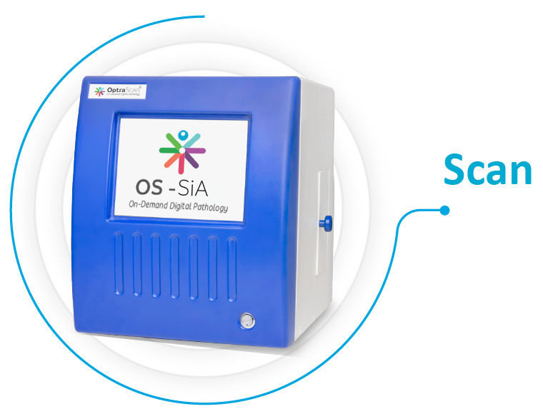
OptraSCAN’s patented OS-SiA™ technology redefines the fundamentals of digital pathology by integrating artificial intelligence directly into the scanning process. OS-SiA™ combines scanning, indexing, and real-time analysis into one device and one seamless process. The technology helps labs deliver faster, more consistent results, without costly infrastructure or additional processing applications or devices. Whether it is a routine diagnostic* lab, regional center, national screening program, or mobile diagnostic* outreach clinic, OS-SiA™ makes screening more efficient, accessible, and actionable, even in low-resource settings with limited on-site expertise.
OS-SiA™ transforms the scanner from a passive image-capture device into an active diagnostic* system. Each scanned slide becomes a source of real-time insights, reducing turnaround time, increasing efficiency, and accelerating clinical decision-making.
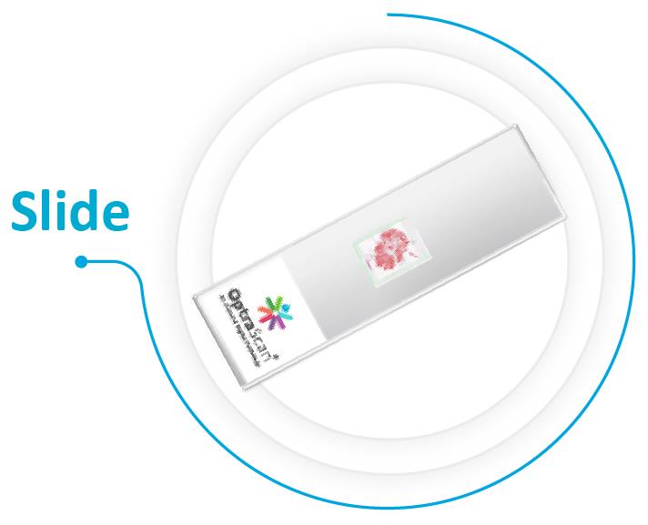
The scanner intelligently interacts with a slide, using 3D Z-stacking and Composite Imaging (OptraSCAN's patented extended-depth-of-field technology). It detects tissue regions, optimizes focus, and combines high-quality images captured across a whole slide in a single plane without losing any data. OS-SiA™ reduces intra- and inter-observer variability.
OS-SiA™ leverages AI to ensure every slide is diagnostically* flawless. Pre-scan integrity check automatically detects physical defects like scratches, dust, and assesses tissue preparation and staining quality. Post-scan image quality assurance validates digital focus, seamless stitching, and optimal color contrast. Finally, it generates a unified diagnostic* QC report, providing pathologists with a guarantee of quality from slide preparation to final diagnosis*, instilling complete confidence in their digital workflow.
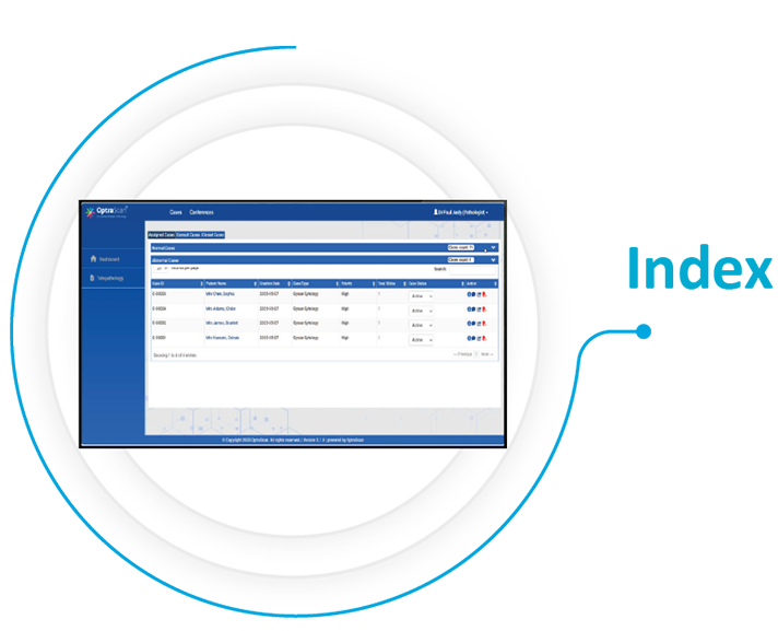
OS-SiA™ automatically generates indexed thumbnails, slide maps, metadata, and contextual information. Once the scan is complete, the slides are already categorized, catalogued, and made searchable. OS-SiA™ instantly tags each scanned region and prepares the database for easy integration into the lab workflow, reducing hours of processing time.
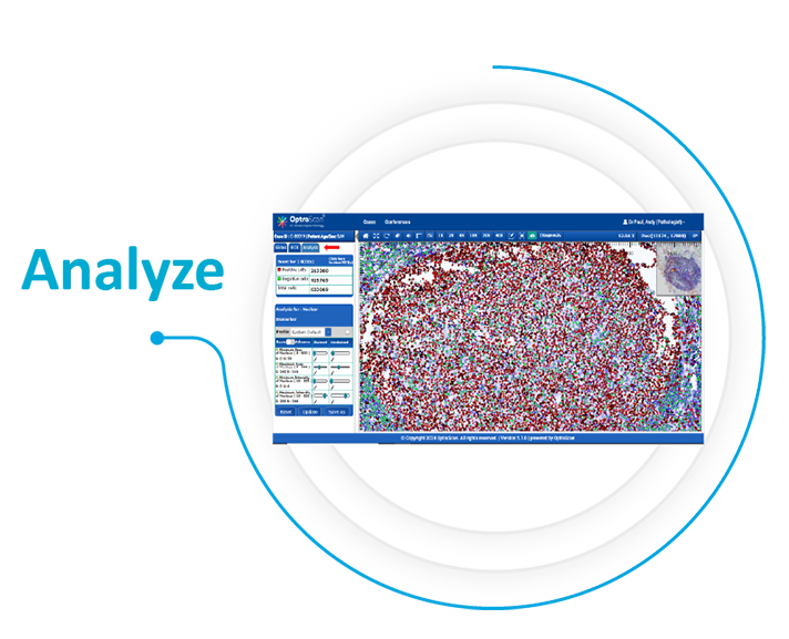
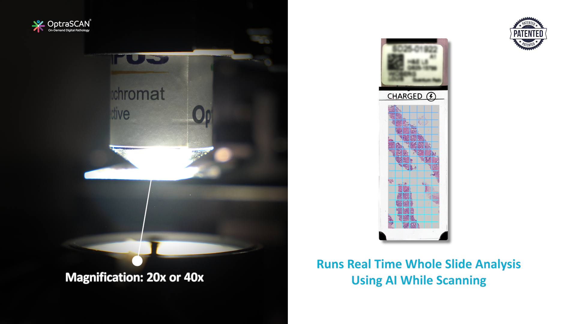
OS-SiA™ runs real-time AI analysis during scanning to provide quantitative insights through morphological measurements, segmentation of normal vs abnormal cases, triaging, and diagnostic* scoring. It provides the turnkey analysis of breast cancer (ER, PR, Ki67, HER2) (TNM staging), lung cancer (PD-L1, EGFR), p53, prostate cancer (Gleason scoring), and cervical cancer cytology (HSIL, LSIL, ASC-H, ASCUS).
By the time the slide is digitized and indexed, the AI analysis is already complete. Pathologists are presented with pre-analyzed intelligence-rich cases, prioritized by urgency and accompanied by AI-generated flags and reports. This slashes diagnostic* turnaround time from hours to minutes.
The final stitched whole slide image (WSI) is immediately and automatically pushed to IMAGEPath® (OptraSCAN’s Image Management System). Anytime anywhere, pathologists can view, pan, zoom, annotate, archive/retrieve the high-resolution small-file-size WSIs and collaborate in real-time, eliminating data storage issues and geographic limitations.

| 1 | Scanning mode: | Brightfield |
| 2 | Magnification: | 20x / 40x |
| 3 | AI assistance: | Real-time analysis with clinically reliable and actionable insights |
| 4 | Image output formats: | JPEG 2000, TIFF, oTIFF, DICOM |
| 5 | Integration: | Bi-directional LIS, PACS, LIMS, HIS via API or HL7 protocol |
| 6 | Architecture: | Cloud-enabled for telepathology and collaboration |

Scanning, indexing, and real-time analysis occur intra-scan (during image capture, not post-scan) in a single step

Works seamlessly with OptraSCAN’s brightfield scanners (OS-SiX, OS-Lite, and OS-Ultra) and IMAGEPath®

Designed for both clinical and research purposesBroad Use Versatility Broad Use Versatility Broad Use Versatility
Eliminates post-processing steps, reduces human error, decreases diagnostic* TAT from hours to minutes, and maximizes throughput

Enhances reproducible results with consistent, AI-driven image quality for every slide every time

Automated workflow handles high-volume caseloads with ease Broad Use Versatility Broad Use Versatility Broad Use Versatility

Requires less investment and minimizes labor, storage, and rescanning costs
OptraSCAN systems are certified under ISO 13485: 2016 and CE-IVD under EU-IVDR 2017/746 for select products

Please fill out the form below and complete all questions with an asterisk (*)