Virtual Immunohistochemistry (IHC) is a revolutionary advancement in the field of digital pathology that combines traditional IHC techniques with cutting-edge imaging technologies. This innovation has transformed the way pathologists evaluate and interpret tissue samples, enhancing diagnostic* accuracy and efficiency.
Virtual staining in digital pathology refers to the use of artificial intelligence (AI) and computational techniques to digitally simulate the appearance of stained tissue samples without physically applying chemical stains like those used in traditional histopathology. This process has the potential to transform workflows, reduce costs, and preserve tissue integrity for further analysis.
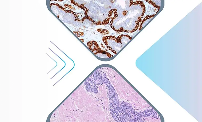
In virtual staining, tissue samples are scanned in their unstained form using advanced imaging techniques such as brightfield or fluorescence microscopy. AI-based algorithms, trained on paired datasets of stained and unstained images, then process the raw image data to digitally replicate the appearance of traditional staining, such as Hematoxylin and Eosin (H&E) or Immunohistochemistry (IHC). The output is a virtual staining overlay that closely resembles a chemically stained sample, allowing pathologists to analyze the tissue digitally without the need for physical staining.
Virtual IHC improves the sensitivity and specificity of diagnostic* tests, providing more accurate assessments of tissue markers and biomarkers. It enables pathologists to analyze slides at a level of detail not possible with traditional light microscopy.
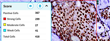
The ability to perform quantitative analysis on IHC staining allows for precise measurements of protein expression, which is essential for determining the aggressiveness of cancers and the response to treatments, efficiency and Workflow Improvement.
Virtual IHC reduces the time spent physically preparing slides and manually analyzing them. With digital tools, pathologists can view and analyze slides remotely, speeding up the diagnostic* process and reducing the risk of human error. OptraSCAN’s automated scanning systems also contribute to this efficiency.
Virtual IHC allows for the use of shared resources across multiple labs, reducing costs and allowing for high-throughput testing in both research and clinical settings.
By enabling the simultaneous detection of multiple biomarkers on the same tissue slide, virtual IHC allows for a more comprehensive understanding of complex biological processes. This is especially useful in cancer research and diagnostics*, where multiple markers need to be analyzed in tandem to understand tumor behavior.
Virtual IHC is not only improving diagnostics* but is also enabling real-time data collection. This is vital in clinical settings, where timely decisions can make a significant difference in patient outcomes. Pathologists can now access high-resolution images of stained tissues instantly, allowing for faster diagnostics* and the ability to consult with peers or specialists in real-time.
In addition, these digital images can be stored and revisited for future analysis, making it easier to track changes in tissue morphology over time. This data can be crucial for ongoing research or for monitoring the progression of diseases like cancer.
Virtual IHC is at the forefront of the next generation of diagnostic* pathology, offering enhanced precision, speed, and efficiency. With the integration of advanced imaging systems like OptraSCAN’s digital pathology solutions and AI-driven analysis, pathologists can expect improved diagnostic* outcomes and more personalized treatment plans. This technological innovation is not just a step forward; it is a leap toward a more accurate and efficient pathology workflow.
By incorporating virtual IHC into your practice, you are investing in the future of pathology—one that delivers better, faster, and more precise results for your patients.
Learn More
Improving diagnostic outcomes through digital workflow adoption.

Everything you need to know about digitizing histopathology workflows.
Where AI stands today and what's needed to close the gap.